CEREBRAL ANGIOGRAPHY
WHAT IS CEREBRAL ANGIOGRAPHY?
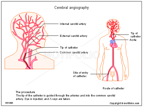
Cerebral Angiography is a minimally invasive medical test that uses x-rays and an iodine-containing contrast material to produce pictures of blood vessels in the brain. In cerebral angiography, a thin plastic tube called a catheter is inserted into an artery in the leg through a small incision in the skin. Using x-ray guidance, the catheter is navigated to the area being examined. Once there, contrast material is injected through the tube and images are captured using ionizing radiation (x-rays). Cerebral angiography is also called Digital Subtraction Angiography (DSA). The images are electronically manipulated so that the overlying bone of the skull, normally obscuring the vessels, is removed from the image resulting in the remaining vessels being clearly seen.
WHAT ARE SOME COMMON USES OF THE PROCEDURE?
Physicians use the procedure to detect or confirm abnormalities within the blood vessels in the brain, including:
- An Aneurysm, a bulge or sac that develops in an artery due to weakness of the arterial wall.
- Atherosclerosis, a narrowing of the arteries or any intraluminal thrombus.
- Total occlusion of vessels (Internal Carotid Artery, Vertbral or Basilar Artery) on MRA/CTA to look for any focal stenosis & need for intervention
- Arteriovenous malformation, a tangle of dilated blood vessels that disrupts normal blood flow in the brain.
- Vasculitis, an inflammation of the blood vessels, generally narrowing them.
- A tear in the wall of an artery, known as a Arterial dissection.
- Acute stroke for intra-arterial TPA or mechanical thrombectomy.
A cerebral angiogram may be performed:
- To evaluate arteries of the head and neck before surgery.
- To provide additional information on abnormalities seen on MRI or CT of the head, such as the blood supply to a tumor.
- To prepare for other medical treatment, such as in the surgical removal of a tumor.
- In preparation for minimally invasive treatment of a vessel abnormality
HOW SHOULD I PREPARE?
- You should report to your doctor all medications that you are taking, including herbal supplements, and if you have any allergies, especially to local anesthetic medications, general anesthesia or to contrast materials containing iodine.
- You will likely be instructed not to eat or drink anything after midnight before your procedure.
- Women should always inform their physician and x-ray technologist if there is any possibility that they are pregnant.
HOW IS THE PROCEDURE PERFORMED?
- In children, cerebral angiography is usually performed with the patient under sedation or general anesthesia.
- Devices to monitor your heart rate and blood pressure will be attached to your body.
- This procedure is often done on an outpatient basis. However, some patients may require admission following the procedure.
- Prior to your procedure, your blood may be tested to determine how well your kidneys are functioning and whether your blood clots normally.
- The area of your body where the catheter is to be inserted will be shaved, sterilized and made numb with a local anesthetic agent. A very small skin incision is made at the site.
- Using x-ray-guidance, a catheter (a long, thin, hollow plastic tube) is inserted into a blood vessel through a tiny hole in the skin made by a needle and directed to the area to be examined. The contrast material is then injected through the catheter.
- A special machine, called a power injector, is used to deliver the contrast material at a precise rate and volume. When the contrast material reaches the blood vessels being examined, several sets of x-rays will be taken. At the end of the procedure, the catheter will be removed and pressure will be applied to stop any bleeding. The opening in the skin is then covered with a dressing. No sutures are needed.
WHAT ARE THE BENEFITS AND RISKS?
BENEFITS
- Angiography may eliminate the need for surgery. If surgery remains necessary, it can be performed more accurately.
- Cerebral angiography presents a very detailed, clear and accurate picture of blood vessels in the brain. This is especially helpful when a surgical procedure or other treatment is being considered.
- Results from cerebral angiography are more accurate than those produced by Carotid Doppler or other noninvasive imaging of the blood vessels.
- Use of a catheter makes it possible to combine diagnosis and treatment in a single procedure.
RISKS
- There is a very slight risk of an allergic reaction if contrast material is injected.
- Women should always inform their physician or x-ray technologist if there is any possibility that they are pregnant.
- If patient has diabetes or kidney disease, low contrast is used to avoid kidney injury.
- Bruising or bleeding at the puncture site, femoral hematoma and infection are few rare side effects
- Although stroke may be a complication associated with cerebral angiography, it is uncommon.